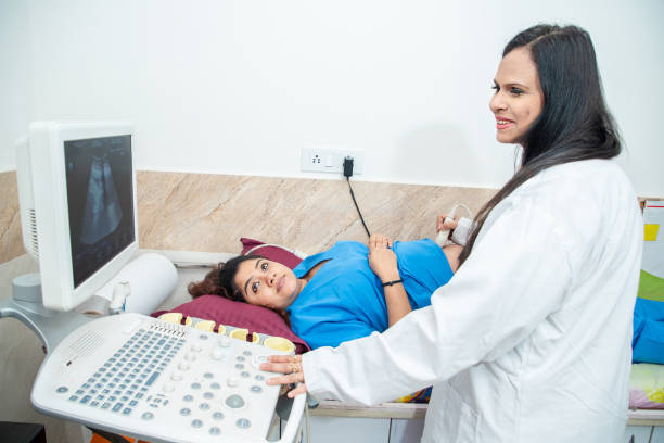
What is Sonography?
Sonography, also known as diagnostic ultrasound, uses sound waves to create images of the inside of your body. It helps doctors visualize internal structures, guiding diagnosis and treatment for various medical conditions.
Ultrasounds are typically non-invasive, with most exams performed using a device outside the body. However, some procedures may require a small device to be inserted into the body for better imaging.

Reasons for Ultrasound
Ultrasound imaging is used for a wide range of purposes, including:
- Monitoring the uterus and ovaries during pregnancy to ensure the baby’s health.
- Diagnosing gallbladder issues.
- Evaluating blood flow.
- Guiding biopsy or tumor treatment procedures.
- Examining breast lumps.
- Assessing the thyroid gland.
- Detecting genital and prostate conditions.
- Checking for joint inflammation, like synovitis.
- Evaluating metabolic bone diseases.
Risks of Ultrasound
Diagnostic ultrasound is a safe procedure that uses low-energy sound waves, with no known risks. However, it does have limitations. Ultrasound is not effective at imaging body parts with gas, such as the lungs or head, or those hidden by bones. In such cases, other imaging methods like CT, MRI scans, or X-rays may be recommended.
Preparation for Ultrasound
Most ultrasound exams require no specific preparation. However, some may have exceptions:
- For a gallbladder ultrasound, you may be instructed to fast for several hours before the exam.
- A pelvic ultrasound may require a full bladder; your healthcare provider will guide you on how much water to drink beforehand and advise you not to urinate until after the exam.
- For young children, additional preparation may be needed. Always check with your healthcare provider for specific instructions.
Clothing and Personal Items
Wear loose-fitting clothing to your ultrasound appointment. You may be asked to remove jewelry from the area being examined and, in some cases, change into a gown. It’s a good idea to leave valuables at home.
During the Ultrasound Procedure
A clear, water-based gel will be applied to your skin over the area being examined. The gel helps eliminate air pockets that could interfere with sound waves. It’s easy to remove and will not stain your clothes.
A sonographer, a trained technician, will use a small hand-held device called a transducer. This device sends sound waves into your body and records the waves that bounce back, creating the images. The sonographer may move the transducer to different areas to get a complete view.
In certain cases, ultrasounds may be performed inside the body through natural openings. Examples include:
- Transesophageal echocardiogram: A transducer is inserted into the esophagus to obtain heart images, usually performed under sedation.
- Transrectal ultrasound: This is used to create images of the prostate by inserting the transducer into the rectum.
- Transvaginal ultrasound: A transducer is inserted into the vagina to examine the uterus and ovaries.
The procedure is usually painless, but you may experience mild discomfort, especially if you have a full bladder or if the transducer is inserted into your body.
An ultrasound typically takes 30 minutes to an hour.
Results of the Ultrasound
After the exam, a radiologist—specialized in interpreting medical images—will analyze the results and send a report to your healthcare provider. Your doctor will then discuss the findings with you.
You can generally resume your normal activities immediately after the procedure.
